case study
Work flow of targeted prostate biopsy in 59 y.o. male with PSA 7.4 ng/ml, no palpable prostate lesion, and prior negative biopsy
Legend:
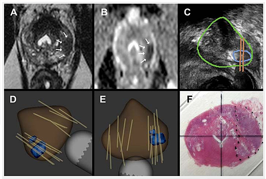
A: T2-weighted axial MRI demonstrating a lesion in the left peripheral prostate.
B: Diffusion weighted MRI showing restricted diffusion (ADC value of 562) within the lesion.
C: Real-time ultrasound image of the lesion (outlined in blue) deriving from MRI fusion in Artemis device.
D and E: 3D reconstruction of prostate, based on ultrasound scan, showing lesion from MRI fusion (in blue) within the model, (D) saggital and (E) transverse views. Tan lines, which are image-captured biopsy sites, show sites of both systematic and targeted biopsy cores. Targeted biopsies in this patient revealed Gleason 7 prostate cancer.
F. Radical prostatectomy specimen showing tumor (dotted line) in whole mount section. Histologically, tumor was a 2 cm Gleason 7 cancer in the left peripheral zone.
Source: UCLA