ARTEMIS –MRI Guided Prostate Biopsy
Incorporating the following features and advantages over existing technology, Artemis provides several imaging enhancements to standard 2D ultrasound.
Automatically converts your 2D monochromatic ultrasound image to an enhanced 3D color image you can then manipulate to plan and manage the patient biopsy process. Greatly increases your ability to examine the prostate for abnormalities or suspicious areas which may need sampling
Advanced needle navigation and tracking you are able to view in real-time
Sophisticated recording of actual biopsy sites sampled; sites can be revisited at any time
View and overlay previous prostate gland volumes and biopsy locations
Artemis accurately assists you in the tracking and management of prostate cancer.
Compatible with all MRI and ultrasound systems
TGA Certified
Provides Rigid and Non- Rigid registration for accurate fusion
Outpatient procedure, left lateral decubitus, periprostatic local anesthetic, standard biopsy gun and needle (No additional supplies)
The only fusion system with a robotic arm, eliminating freehand movement error
Advanced proprietary motion compensation technology corrects patient movement during biopsy
Real time, automatic, live fusion
Whole prostate gland sampling, with patented needle tracking and recording
Re-sample the same sites previously biopsied for active surveillance
Send MRI data directly to Artemis for targeted biopsy via cloud
Send biopsy data and Pathology results from Artemis to ProFuse for possible treatment planning
Artemis generate patient report with a colour 3D fusion image of the prostate and regions of interest
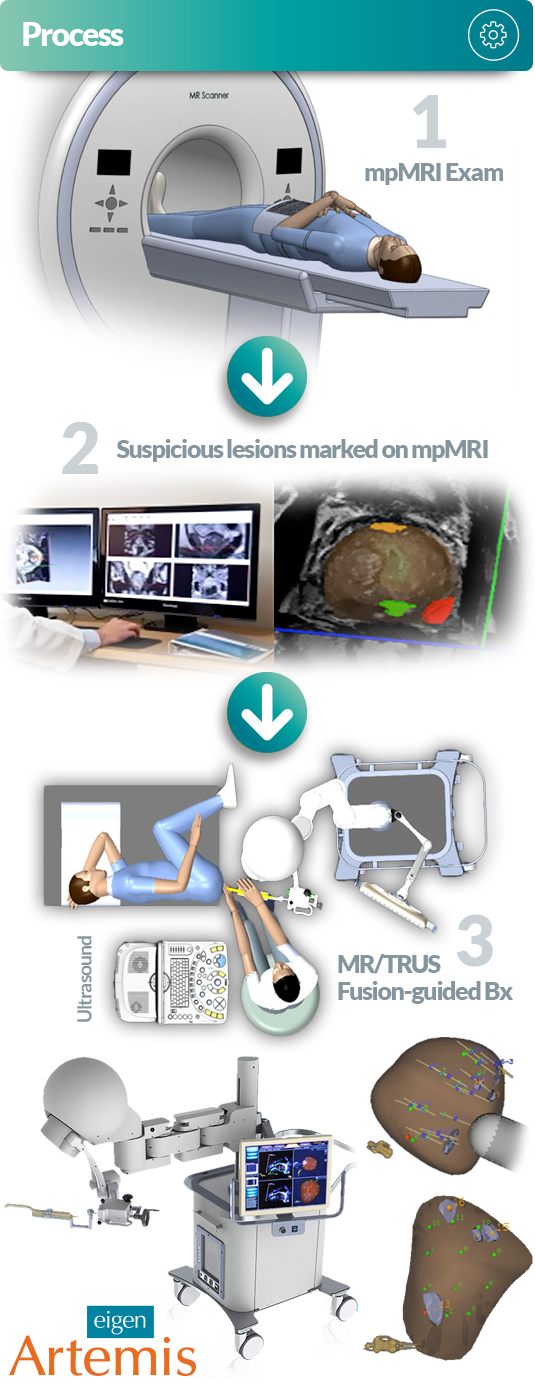
Artemis – Smart Prostate Biopsy Steps
The first step is for your doctor to take a 360° scan of your prostate using a standard 2D ultrasound system connected to Artemis. Next, Artemis automatically converts the standard 2D ultrasound image into an enhanced 3D prostate image. Your physician can easily adjust the prostate boundary and contour to customize the image even further.
The result is a clear 3D prostate rendering that provides full 3D rotational functionality along with real-time views of the prostate in multiple planes – coronal (top), sagittal (side) and transverse (front). The visualization experience is amazing, allowing your doctors to see the entire prostate as one clear complete image. The power of 3D helps to increase your physician’s ability to examine the prostate for abnormalities or suspicious areas that might otherwise not be detected. By seeing the gland in 3D it’s easier for your doctor to be sure the entire prostate is examined correctly. Likewise, it’s easy to find any areas of potential concern.
Artemis and MRI (An Optional Scanning Module)
The Magnetic resonance imaging (MRI) Fusion Module allows physicians to integrate three images (MRI, 3D-Artemis rendering, and real-time ultrasound images) into one single visual.
MRI is a test that uses a magnetic field and pulses of radio wave energy to make pictures of organs and structures inside the body. In many cases MRI gives different information about structures in the body than can be seen with an ultrasound. MRI also may show problems that cannot be seen with other imaging methods.
The fusion function gives physicians the most sophisticated imagery and navigational ability available. It provides high resolution along with exact structural dimensional functionality. The result is a significantly enhanced prostate examination and biopsy sampling. Your physician now can have the confidence of precise needle placement in areas identified by the radiologist during the MRI scan. The MRI fusion feature allows physicians to scale an exam and/or biopsy – from the simplest to the most advanced procedure – to meet patient needs. MRI fusion is typically useful in patients who require repeat biopsies or for patients with a negative biopsy and continued PSA escalation. MRI fusion is also helpful in patients with specific areas (or lesions) of concern of for patients with very low-risk or very high-risk disease. The optional fusion functionality allows your physician the ability to customize each procedure as needed. And it helps to simplify a busy practice by having one system that can handle every type of prostate exam and biopsy.
Step three is navigation and recording. Artemis allows physicians to achieve exact needle placement by using the system’s real-time dynamic needle track navigation capabilities. This unique software tracks the needle as the physician glides the ultrasound probe along each biopsy template. The software lets physicians precisely navigate where they want to take the sample by using Artemis’s XYZ navigation guidance process (much like a video game or GPS). Once the doctor aligns the ultrasound probe with the 3D template image, the probe can be temporarily locked in place and a biopsy sample is taken. Artemis then precisely records the location of the biopsy sample site for reporting. This unique software–hardware interface makes Artemis easy to learn and simple to use. Best of all, the tracking function give doctors the ability to revisit any and all previous biopsy sample sites. The process helps to eliminate blind-biopsies and guessing where the needle is.
If a patient has had a previous biopsy with Artemis, his physician can access the biopsy recording for repeat biopsy planning, including overlaying a previous prostate volume and actual biopsy site locations. This gives your doctor the ability to re-sample the same biopsy sites, sample different sites, or saturate a particular area. Most importantly, the tracking function can be used to conduct precise serial biopsy sampling for “Active Surveillance” patients. This important function can also help physicians manage patients who had a previous biopsy and patients who have already been treated for prostate cancer. Essentially, Artemis helps to create a long-term patient relationship by providing a sophisticated biopsy management platform that helps ensure that patients who return to the same clinic have their history of accurate biopsies locations recorded.